|
My
own Pedigree
News Our
Dogs Shows Beachwalks
Our
other Passion
|
What
is now a wrong colour? A
wrong colour is a
not desired colour, which is
not acknowledged by the
Council of Management
and the FCI
( Federation Cynologic International
)
and is not described in the race standard of the race association of
the
NNFC (Dutch Newfoundland Club). Through new sciences study's of the last 5-year: are through the "University of Saskatcheawan Department of Animal and Poultry Science by Professor Dr. Mrs. Sheila Schmutz" are based on DNA and phenotypic comparisons other conclusions regarding the genes of the A locus B , C , E , S and a K locus have been added. In the old situation it was : Thus
therefore the genes lie which stipulate the colour of the coat, as far
as I know, on
9 loci
: A,B,C,D,E,G,M,S,t. Well the new situation is now : For
instance, the genes
that determine
coat color,
to my knowledge,
at 10
loci
:
a,
KB,
B,
C, D,
E, G, M, S,
t. On a locus
"a place on the DNA string"
is only room for
Maximun two genes, "of every parent one
for each gene" . The
colour code is in homozygous form
(pure) for the
black =
KBKBBBCCDDEESStt.
Dark
pigment (
eumelanin) is black and
brown, yellow
and
red are light
pigments (phaeomelanin). §
at
gene : is standing for black/liver and tan. §
KB
gene : is standing for dominance (blac
K = Solid) dark pigment over the whole body §
kbr
gene :
§
b
gene : is standing for brown.
Three different mutations in this gene (
bs,bd,bc )
can
produce brown. §
C
gene : complete pigmentation, gives all deep red and brown races.
§
D
gene : is standing for intensive pigmentation §
d
gene : is standing for
dilution §
E
gene : is standing for dark pigment (
eumelanin
) concerning the coat
§
S
gene : is standing for divided stopping the white colour, except
sometimes a white mark on the chest and a couple of white toes §
si
gene : stands
for Irish
spotting
"pattern
or UnderSides
or Pseudo-Irish
Spotting"
is a white spot
on his chest, white feet
and tailtip,
sometimes with
some white on the
muzzle in a more
or less symmetrical
pattern. §
sp
gene :
piebald spotting, a large white mark or white
without pattern.
§
T
gene : is standing for Ticking §
t
gene : is standing for reducing Ticking.
The
key to understanding dog genetics is simply this: there are two types
of pigment which create coat colour in dogs (and most other mammals).
Pigment
is just the thing that gives each strand of hair its colour, just like
pigment in paint or dye, or pigment in your own hair.
EUMELANIN Eumelanin
is black pigment. All
black areas on a dog are caused by cells producing eumelanin. However,
there are genes which turn eumelanin into other colours - liver (brown), blue
(grey), or isabella (a dusty pale brown) If
a dog has any of the genes to turn its black eumelanin into liver, blue or
isabella then all of the black in its coat will be changed. This
is because these genes restrict and/or alter the production of eumelanin,
so the cells aren't able to produce full-strength pigment. We
call blue and isabella dogs "dilutes" for this
reason. As
well as being found in the coat, eumelanin is present in the other parts
of the dog that need colour - most notably the eyes (irises) and nose.
When
we talk of dogs that are "black pigmented ", "liver
pigmented", etc, we mean that is the colour of eumelanin that the dog
can produce. Sometimes
these dogs have no eumelanin at all in their coats ( their skin cells
produce only the other type of pigment, phaeomelanin),
PHAEOMELANIN The
second type of pigment, less important than
eumelanin, is phaeomelanin. This
is red pigment. Phaeomelanin
is produced only in the coat .
So far so good, but this doesn't seem to explain all the coat colours in dogs - how about white ? WHITE White
is not
really a
color,
so white
hairs on animals
is not caused by
pigment, but
a lack
of pigment. It
is a lack
of both eumelanin
and phaeomelanin.
White areas on
animals are
simply caused
by the pigment
cells, there
is no creation.
Sometimes the entire
animal affected
by e/e-gene
(read Canadian
Shepherd),
or as in albinos
( ca
gene)
where no pigment
is present and
sometimes just
parts of the dog
without pigment
is influenced,
like dogs
with white
markings where
si
and sp
gene responsible.
It can affect
the production
of eumelanin
in the eyes
and forenose
which nose
pink
and the
eyes to
blue change
(or red in
the right albinos).
This type of
white has no
effect on eumelanin,
so any black
/ brown
/ blue
/ isabella
areas on the
coat will
remain pure, and
the eyes and forenose, paw
pads will
be too.
Research
has shown that a recessive e
allele
at the Extension (E)
gene is at least partially
responsible for cream and white coat color by
the Canadian Shepherd. The
(E)
gene, now
identified as the Melanocortin 1 receptor (MC1R) gene,
is one of the two genes known to code for alleles that are absolutely
fundamental to the formation of all German Shepherd Dog colored coat
variations. When the recessive
allele is inherited from
each
breeding pair parent, the e/e
genotype offspring of certain breeds, including white coat dogs from
German Shepherd breed lines,
DISTRIBUTION OF PIGMENT The colour genes in dogs do two things - they determine the eumelanin and phaeomelanin colours/shades, and they control the distribution of these two pigments. They tell certain cells to produce eumelanin, others to produce phaeomelanin, and sometimes they tell them to not produce pigment at all. Exactly which cells are told to produce what is determined by the exact set of genes, although it can be random to a certain degree (eg puppies may have slightly different white markings to their parents, or patches in different places). Sometimes genes can even tell cells to switch which type of pigment they are producing every once in a while. This means that as a hair grows, it becomes banded with black and red, because the cell produces black (eumelanin) for a while, then changes to red (phaeomelanin), then back to black, etc. It's a bit like when you highlight your hair and after a while the roots start to show through. The overall colour of an animal with this sort of black and red banding will generally be a muddy brown from a distance, and close up you will be able to see the black parts of the hairs. It's called agouti (aw-gene) , and it's the colour of wild rabbits and mice, as well as a large amount of other mammals. It's popular amongst wild animals because it provides very good camouflage. It also occurs in dogs, but it looks a bit different (like the colour of a wild wolf rather than a rabbit) and isn't very common. All of this probably seems a bit confusing at this point. Many genetics sites gloss over the types of pigment for precisely this reason. However, I've decided to include this information because it really is crucial to your understanding of dog genetics (and the genetics of other animals too). Once you've grasped the idea of the two types of pigment, and understand how they work, the rest should be plain sailing.
The
yellow does not have been confused with
small
e-gene, like the
Labrador,
in homozygous form (ee) gives this a junction of a black and a brown
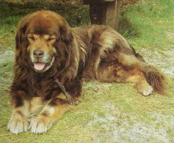
A
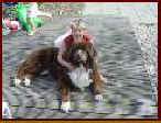
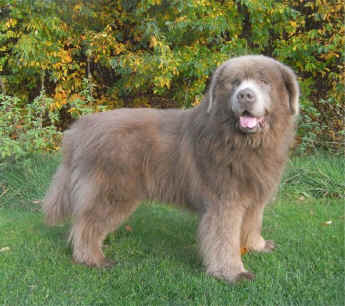
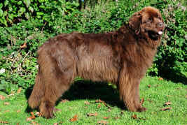
brown Newfoundlander (si x
si-gen) with white
"Irish spotting" (code
KBKBbbCCDDEEsisitt) (see
photograph). §
Code
KBKBbbCCDDEEspsptt
(wrong colour) (see
photograph). §
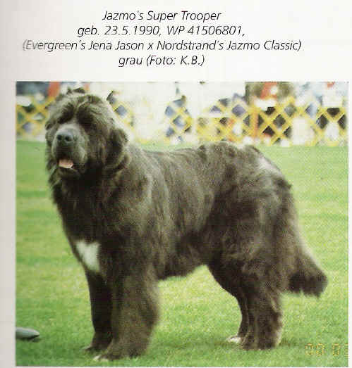
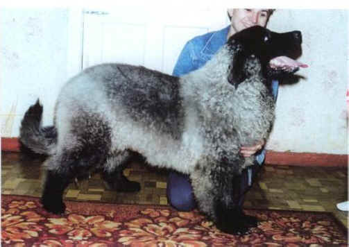
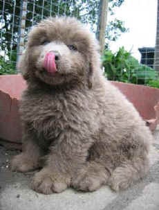
Code
KBKBBBCCddEESStt = grey with black nose capsule, and foot soles and lips
(wrong colour) (see
photograph). §
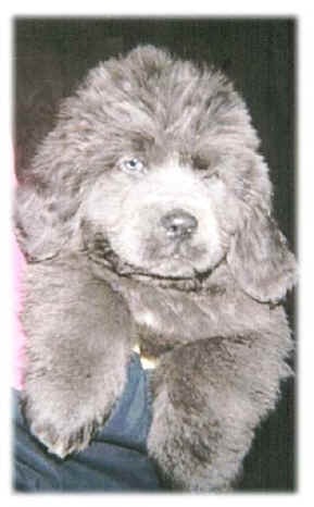
Code
KBKBbbCCddEESStt = grey with brown nose capsule and foot soles and lips
(wrong colour) (see
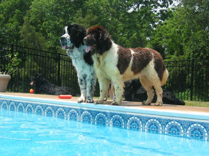
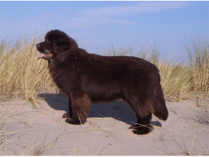
photograph). Is
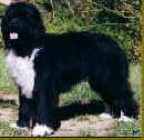
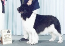
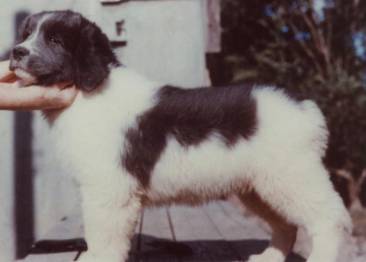
it now a white-black or black-white Newfoundlander (see
photograph)? The
proposition that the piebald "sp-gene" is not responsible for
a white mark, but for a white dog with coloured marks. First,
even for all
clarity, the code:
KBKBBBCCDDEEspsptt
, therefore in my opinion a
black-white Newfoundlander and
I will try to explain why
I think so.
S-locus: S, si, sp S = stop of white,
si, sp, are genes which
brings white in order of
dominance, therefore everybody talks about a
white factor and
white is
NO COLOUR but the cause that these genes the
pigment widen-honoured which is in a dog present, as above already
mentioned. Everybody
is talking about a
black dog with
no pigment
mark "so white". sp-gene
do that
without
pattern, in fact random white
marks are falling in the coat. The
genes on
the S-locus
are not
complementary
to other genes
in the code
such as the
KBB/b/D
locus and
the Ee
locus, such
as locks and
keys or
Yin /
Yang and can
move independently of the
other genes
between the code and manifest
themselves "as
exemble" as
a dominant
dog :
" KBKBBBCCDDEESsitt
"
but carriers
of the
white
-
si
or sp
factors. NOTE:
Some of these alleles at the S locus may have incomplete
or co-dominance. There may be genes independently modify (modifications change a part of an assignment, so the interpretation and implementation differ) from the original manner specified place for example in the SS-genes (white patch) or a black and white litter (spsp-genes) where there is not one in the same pigmentation and the Irish Spotting marks (sisi-genes) is one white paw can be more white than the other instance. And
Ticking -TT-gene - gives therefore mark colours
in the white and
tt-gene hold back
this. If
you
cross therefore
two
black dogs with the white inherit factor, the
recessive
sp-gene =
KBKBBBCCDDEESsptt
x
KBKBBBCCDDEESsptt
you get : §
25%
black =
SS pure breed §
50%
black =
Ssp impure breed §
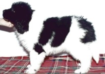
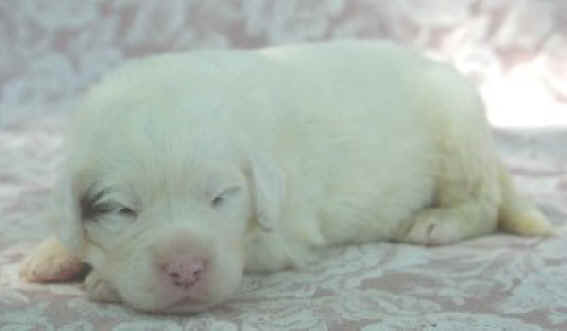
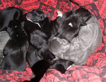
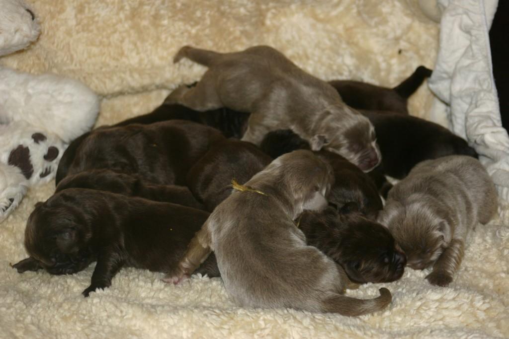
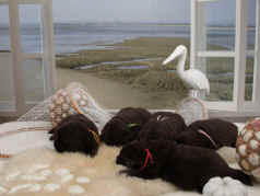
25%
fur =
spsp pure breed for
fur, without fixed pattern (see
photograph) §
A
cross of two
furs from the white factor spsp x
spsp = 100%
fur without fixed pattern §
Two
black with white mark and white tail point, feet and white chest mark:
sisi x
sisi =
100% have this property in
more or less in the
symmetrical pattern §
Two
black with the Ssi x
Ssi = 25% blacks
SS pure, 50% are black recessive
Ssi
impure, 25% sisi
pure
black with white marks
Take
a brown /
grey
dog
with the dd
gene (wrong
colorr)
X a
clear genotypical dominance
brown dog
= SS
I
want to
explain
the behaviour of the travelling of the genes of the white
si-gene
"Irish Spotting" what is travelling
for generations and is coming out if both parents are carrier (= 25% who has
it). Yes it is also
travelling even after using dominant black
dogs ( KBKBBBCCDDEESsitt
) except with dogs who are §
Did
you know that all colour code combinations of the Newfoundlander in pure
breed and impure breed (homozygoot: KBKBBBCCDDEESStt
and heterozygote "collective
code" : ayatKBkbrkyBbCcaDdEESsispTt
amounts, so far as I can see, more than a 500. §
This
way there is still a couple wrong colour codes. § Code ayayKBKBBBCCDDEESStt, is a black-yellow/brown (agouti Fawn) pigment dog "looks like a brown one"with a black forenose and mirror/mask. §
Code
ayatKBKBCCDDEESStt, is a black yellow/brown
=
agouiti Fawn pigment dog "looks like a brown
one "with a black forenose and mirror, but by heterozygote form because the
ay-gene are dominance over the
at-gene. §
This
one with the small b for brown on the locus
ayatKBKBbbCCDDEESStt, therefore a
liver colour with more yellow "looks like a yellow dog" with a
liver coloured nose capsule and mirror. §
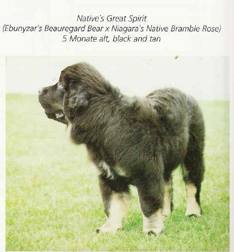
Code
atatkykyBBCCDDEESStt, is for
Black and Tan "brown-yellow colouring and
looks like crème colour to halfway the legs and/or the mask
" §
Code
atatkykybbCCDDEESStt, is for
liver and Tan "brown-yellow colouring at
brown, looks like "crème-grey" also called “sandy” on the
legs and/or the mask" (see
photograph). §
Code
KBKBBBCCDDEEspspTt, is white-black with Ticking (heterozygote
“impure”
form). §
Code
KBKBBBCCDDEEspspTT, is white-black with Ticking (homozygote “pure”
form
However, the indication is that black/liver only shows themselves in homozygote “pure” form, therefore both parents are carrier of the at-gene, ( is also present by the Tibet Dog, see also , “ Pre history / Primal Source /Tibet Dog" By
strong
INBREEDING there is a great risk in the future to get back this
wrong colour, especially with combinations of the
K
= short hair =
dominance k
= long hair F
= phenotypic calculation of the Augustine monk
Gregor Johan Mendel
(Czechia
1822 --.1884). By
means of
F1-generation becomes 100% short hair, therefore
K x
k = 100%
short hair =
Kk. In
the
F2 generation becomes the
KkxKk = 75%
short hair ( of which, 25%
KK
is pure breed, 50%
impure breed Kk,
Kk )
and 25% long hair
kk homozygoot
pure breed.
( 3: 1 )
. Conclusion:
Long hair x long hair = is therefore
ALWAYS long
hair.
Tyrosinase
Related Protein 1 (TYRP1) is the gene responsible for brown coat colors in
dogs (and mice and cattle and cats).
The standard color of the eyes is
brown for dogs.
Occasionally
have dogs
with black
pigment amber
eyes, Note : The
first gene that causes at least some
spotting patterns in dogs was
identified and published in 2007. MITF has been shown to cause spotting of the piebald or random type in crossbreds by Rothschild and colleagues. It also caused the Landseer pattern in Newfoundlands. However the causative mutation was not identified in their study. Another study
presented by Karlsson on behalf of the Broad Institute and the
Univerisity in Uppsala, Sweden focussed primarily on Boxers. They have
shown that MITF is the gene causing the
solid, flashy, and virtually white forms in Boxers and Bull Terriers.
They suggest that two mutations in the promoter region of the MITF form
that works in the pigmenation pathway (MITF-M)
are necessary for white in these breeds. One of these is called a SINE
(short interspersed nucleotide element) and the other
is a string of repeated alleles that
varies in length in different spotted dogs, which they call a "Length
Polymorphism or LP". Their data
suggest that the LP is found to be longer in breeds with white markings
than in dogs with no white. White Undersides or Irish Pattern Clarence C. Little (1957) discusses the "pseudo-Irish Spotting" phenotype in the flashy Boxer and then raises the perplexing pattern he refers to as "Irish spotting", in dogs such as Basenji. He also suggests that plus and minus modifiers are likely. He says the si causes this pattern and that is recessive to the S allele for solid, but dominant to the allele causing piebald spotting (sp). MITF and White Spotting in Dogs: A Population StudyThis study was designed to determine if one of the variants found in our laboratory, or previously reported in microphthalmia-associated transcription factor (MITF), was associated with one or more spotting patterns in dogs. None of the rare variants found in the coding sequence consistently occurred in dogs of any particular spotting pattern. However, an insertion of a short interspersed nucleotide element (SINE) over 3000 bp 5′ of the MITF-M start codon (Karlsson et al. 2007) did fit with random spotting in many dog breeds. Most (319/324) dogs of 45 breeds fit 1 of 2 inheritance patterns. All dogs that were homozygous for the SINE had white markings that either covered at least the ventral surface (mantle pattern) or most of the body (piebald or extreme white spotting). In most breeds, dogs heterozygous for the SINE insertion were solid colored or had minimal white, such as on the toes, but in some others, heterozygotes had white undersides, often with a white collar in the pattern called pseudo-Irish by mr Clarence Charles Little (1957). However, none of the 15 dogs of 5 breeds in which all individuals have markings known as Irish spotting had the SINE insertion. Finally, we studied RNA expression in skin. The 2 MITF-M forms, M+ that contains an extra 18 bp that adds 6 amino acids between exons 5 and 6 and the M− form, were present. MITF-M is considered to be specific to melanocytes but was found in skin from a white Samoyed. A putative pseudogene containing exon 1M was also identified. As a resource
for building a dog genetic map and as a tool to study the genes
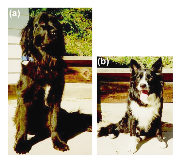
responsible for behavioral and morphological differences in the dog, an intercross
was created between a male Border Collie (b) and
a female Newfoundland (a). The
Newfoundland parent had a small patch of white on the chest and was
otherwise completely black. The Border Collie used in this cross had
markings characteristic for the breed - black with
white markings on the face, chest, neck, tail tip, ventral
abdomen, all four digits, and extending up the front legs to the carpals.
These markings have many similarities to the white spotting patterns of
other mammals. The Border Collie's sire and dam had the same markings,
consistent with homozygosity for the causative loci. This cross provides
the opportunity for analyzing the inheritance of the white spotting
pattern exhibited by the Border Collie. Six F1
animals were produced which had medium-sized white
patches on their chests. These six dogs were intercrossed
to produce 25 of F2 progeny.
In the F2 generation,
7
out of 25
had markings like the Border Collie parent,
consistent with the phenotype being caused by a recessive allele
of a single locus.
The SINE
insertion 5′ of MITF-M first described by Karlsson
et al. (2007) was associated with white markings in many and
diverse
breeds in this study, suggesting that it is an “old”
mutation. There is considerable debate about the age of
particular breeds. Science Links about this issue are : Undersides or Pseudo Irish Spotting (2000) MITF and White Spotting in Dogs: A Population Study (2009) Prof. Dr. Sheila M. Schmutz University of Saskatchewan Saskatoon, Canada S7N 5A8 updated in january 9, 2012 |
|||||||||||||||||||
